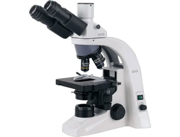
Microscopy has revolutionized biological research by allowing scientists to observe structures beyond the limits of the naked eye. One of the most effective techniques for visualizing biological samples with intricate details is Polarized Light Microscopy (PLM)
polarized Light
Polarized light consists of light waves that vibrate in a single plane rather than multiple directions. This controlled light behavior is key to improving contrast in microscopy.
How PLM Works
PLM uses polarized light to interact with anisotropic materials—substances with different properties in different directions—allowing scientists to see otherwise invisible details.
Components of a Polarized Light Microscope
A PLM setup includes:
- A light source
- Polarizers (to filter light)
- An analyzer (to detect polarized light changes)
- High-resolution optics
The Science Behind Polarized Light
Properties of Light Waves
Light consists of waves oscillating in multiple planes. Polarization restricts these oscillations to a single direction, improving image clarity.
Birefringence and Its Role in Microscopy
Birefringence occurs when light passes through materials with different refractive indices in different orientations. Many biological samples exhibit birefringence, making them ideal for PLM.
Interaction of Light with Biological Structures
Polarized light interacts uniquely with fibers, proteins, and crystalline structures, revealing intricate details not seen in standard light microscope
Advantages of Polarized Light Microscopy
- Enhanced Contrast: Highlights birefringent structures in biological tissues.
- Improved Visualization: Detects subtle differences in sample composition.
- Non-Invasive Technique: Preserves delicate biological structures.
Applications of PLM in Biological Studies
- Muscle Fibers & Collagen: Identifies fibrous structures and connective tissues.
- Crystals in Tissues: Detects uric acid crystals in gout studies.
- Plant Cell Walls: Examines cellulose organization in plants.
Comparing PLM with Other Microscopy Techniques
| Feature | PLM | Brightfield | Fluorescence | Electron |
| Contrast | High | Low | Moderate | High |
| Resolution | Moderate | Moderate | High | Very High |
| Sample Type | Birefringent | Transparent | Fluorescent | Electron-dense |